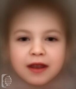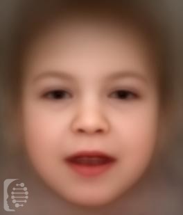
What is Mucolipidosis Type IV?
Mucolipidosis Type IV is a rare genetic condition first identified in 1974. Between then and 2010 only around 70 cases have been diagnosed.
70% of the cases of the syndrome are in individuals of Ashkenazi Jewish ancestry.
This lysosomal storage syndrome is metabolic and progressive. Characteristic features of the syndrome include delayed psychomotor development and visual impairment.
Syndrome Synonyms:
Ml IV; Ml4; ML4; MLIV; Sialolipidosis
What gene change causes Mucolipidosis Type IV?
Mutations in the MCOLN1 gene are responsible for the syndrome. It is inherited in an autosomal recessive pattern.
Autosomal recessive inheritance means an affected individual receives one copy of a mutated gene from each of their parents, giving them two copies of a mutated gene. Parents, who carry only one copy of the gene mutation will not generally show any symptoms, but have a 25% chance of passing the copies of the gene mutations onto each of their children.
The syndrome is a lysosomal storage disease caused by a deficiency of arylsulfatase B (ASB).
What are the main symptoms of Mucolipidosis Type IV?
Typical Mucolipidosis Type IV is a severe form of the disorder and can cause serious symptoms. The atypical form of the disorder is much less severe and symptoms can vary widely between individuals.
Symptoms generally appear leading to a diagnosis in infancy, depending on their severity. These symptoms include poor muscular and mental coordination and reduced muscle tone.
Physical characteristics of the disorder include clouding of the cornea, retinal degeneration, crossed eyes, puffy eyelids, myopia and a hyper sensitivity to light.
Possible clinical traits/features:
Spastic tetraplegia, Progressive retinal degeneration, Strabismus, Infantile onset, Microcephaly, Genu recurvatum, Gait disturbance, Cognitive impairment, Incoordination, Hyperreflexia, Opacification of the corneal stroma, Abnormality of mucopolysaccharide metabolism, Abnormality of abdomen morphology, Cerebral dysmyelination, Cerebellar atrophy, Babinski sign, Abnormality of retinal pigmentation, Aplasia/Hypoplasia of the abdominal wall musculature, Absent speech, Palmoplantar keratoderma, Autosomal recessive inheritance, Nystagmus, Optic atrophy, Retinopathy, Photophobia, Everted lower lip vermilion, EEG abnormality, Decreased light- and dark-adapted electroretinogram amplitude, Developmental stagnation, Coarse facial features, Dystonia, Dysplastic corpus callosum, Intellectual disability, Muscular hypotonia, Abnormal electroretinogram, Abnormal nasal morphology, Ganglioside accumulation, Narrow forehead, Neurological speech impairment, Microdontia.
How is it diagnosed?
To find out if someone has a diagnosis of Mucolipidosis Type IV, it is important to have a consultation and evaluation with a clinical genetic specialist. Specialists may also suggest specific genetic testing or other types of tests to help reach a diagnosis. FDNA’s AI technology can help speed up the diagnostic process by analyzing facial features and other health information.